 Translate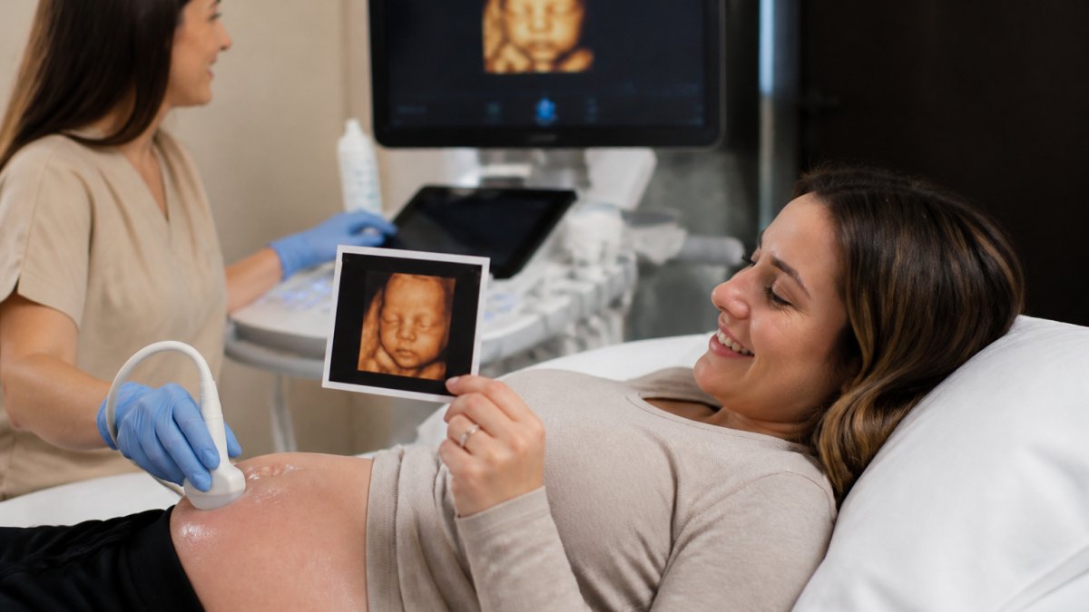
Table of Contents
- Why the Best Time for a 3D Ultrasound Falls Between 24 and 32 Weeks
- What Week Is Best for a 3D Ultrasound? A Trimester-by-Trimester Breakdown
- 3D Ultrasound Preparation Tips That Improve Image Clarity
- What to Expect at a 3D Ultrasound Appointment
- How to Get Baby to Move for an Ultrasound
- Factors That Affect 3D Ultrasound Image Quality
- Medical vs. Elective 3D Ultrasounds: What's the Difference?
- Frequently Asked Questions
Last Updated: September 28, 2026
Why the Best Time for a 3D Ultrasound Falls Between 24 and 32 Weeks
The best time for a 3D ultrasound is between 24 and 32 weeks of pregnancy, when you get the clearest view of your baby's face, hands, and tiny features.
Here's what's happening in those weeks. Your baby is putting on subcutaneous fat, the layer under the skin that gives a face its round shape. Before 24 weeks, that fat is thin. The face looks bony and sharp. After 32 weeks, your baby is bigger and curled up tight. The amniotic fluid that acts as a window for the sound waves starts to drop.
That fluid is your camera lens. Less fluid means a fuzzier picture.
The sweet spot sits in the middle: enough fat for a soft, full face and enough fluid to float your baby into view.
The American College of Obstetricians and Gynecologists guidance on ultrasound in pregnancy notes that ultrasound is a standard part of prenatal care, though the timing of each scan serves a different purpose. A keepsake scan is a different animal from a diagnostic one, and we'll get to that.
Below, we'll show you what each week looks like and how to prep for the clearest images.
What Week Is Best for a 3D Ultrasound? A Trimester-by-Trimester Breakdown
Weeks 28 to 32 are the single best stretch for most parents. The face is full, the fluid is still generous, and your baby has room to turn.
But the "best" week depends on what you want to see. Let's break it down by trimester.
Second Trimester: Weeks 24 to 27
Weeks 24 to 27 land in the second trimester. Your baby's facial features are formed and the fat layer is building.
You'll often catch a full profile here, with nose, lips, and chin reading clearly, and the baby still has space to stretch and turn.
The trade-off? The face can look a little lean. Cheeks fill in later.
Third Trimester: Weeks 28 to 32
Weeks 28 to 32 sit in the third trimester, and this is where the magic happens. The cheeks are plump. The fat pads around the eyes and mouth are in place.
This is the range most first-time parents target, and it's the range we recommend for keepsake images.
| Timing | What You'll See | Best For |
|---|---|---|
| Weeks 24-27 | Full profile, leaner face | Early bonding, gender confirmation |
| Weeks 28-32 | Round cheeks, full features | Keepsake images, gender reveal |
| After 32 weeks | Tighter space, less fluid | Limited; face may be hidden |
3D Ultrasound Preparation Tips That Improve Image Clarity
Good images start before you ever lie down. Most providers offer advice on preparation. Here is a plan to help you prepare for your 3D ultrasound.
The 48-Hour Hydration Schedule
Amniotic fluid is your camera lens: more of it gives sound waves a cleaner path to your baby's face, and hydration is one of the few variables you control. Parents who hydrate steadily for two days tend to get sharper images than those who chug a bottle in the parking lot.
- Two days out: aim for roughly 8 to 10 cups of water spread across the day, not slammed at once. Steady intake is what your body uses to maintain fluid volume.
- The day before: keep the same pace. Add water-rich foods, watermelon, cucumber, oranges, soup, which hydrate without forcing you to drink past comfort.
- One hour before your appointment: drink about 8 to 12 ounces. Enough to top off, not enough to make you desperate for the restroom mid-scan.
- Skip the giant pre-scan jug. A sudden flood mostly fills your bladder, not your amniotic sac, and it makes the session uncomfortable.
Eating for a Cooperative Baby
Fetal activity is driven partly by your blood sugar. A light snack 30 to 60 minutes before the scan, with a little protein and carbohydrate, keeps a baby active without sending them into a frenzy. A wildly thrashing baby is as hard to photograph as a sleeping one, so the goal is alert, not wired.
Day-Of Checklist
- Wear a two-piece outfit so your belly is easy to access
- Skip lotions, oils, and self-tanner on your abdomen, they interfere with probe contact
- Bring your partner and any kids you want in the room
- Arrive a few minutes early so you are not rushed onto the table
- Pad your schedule by at least an hour in case your baby needs coaxing
What Prep Cannot Fix
No amount of water overcomes a baby who has wedged their face against the placenta or turned toward your spine. Prep stacks the odds in your favor but does not control the outcome, which is why a good provider builds in time for a second attempt.
What to Expect at a 3D Ultrasound Appointment
A 3D ultrasound appointment is a relaxed, non-clinical session. You lie back, a sonographer moves a transabdominal probe over your belly, and images appear on a screen in real time.

Here's the typical flow:
- You check in and get settled on a padded recliner
- The sonographer applies gel and starts the scan
- You watch your baby move on the monitor
- The tech captures still images and short video clips
- You pick your favorites for keepsakes
How to Get Baby to Move for an Ultrasound
An uncooperative baby is the number one reason for a repeat scan. The fix is usually simple movement on your part.
Try these, in order:
- Change positions: roll to your side, then your back
- Walk around the room for a few minutes
- Drink something cold and wait
- Gently press or jiggle your belly if the tech says it's fine
- Eat a small snack for a quick energy bump
Factors That Affect 3D Ultrasound Image Quality
Image clarity is not one variable, it is a stack. Week of pregnancy sets the ceiling, but several other factors decide whether you reach it. Understanding each mechanism tells you what you can influence and what you cannot.
Fetal Position: The Biggest Single Factor
Sound waves travel in a straight line and cannot see through bone. A baby facing your spine presents the back of the skull, and no machine can photograph a face through that. A baby facing outward, a favorable position, gives you the shot.
Amniotic Fluid Volume: Your Acoustic Window
Fluid is the medium sound waves pass through to reach your baby. More fluid means a wider, cleaner path; less fluid means the waves scatter and the image softens. Fluid volume peaks in the late second trimester and declines as pregnancy progresses, which is one reason the 28-to-32-week window outperforms later dates.
Maternal Body Habitus and Penetration Depth
This is the topic most consumer articles skip, and one of the most searched. Ultrasound waves lose energy as they travel through tissue, so more tissue between the probe and the baby means weaker waves and softer clarity, the same principle that makes it harder to hear someone through a thick wall than a thin one.
Placental Location
An anterior placenta, attached to the front wall of the uterus, sits between the probe and your baby and can shadow part of the view, particularly facial images. A posterior placenta, on the back wall, leaves a clearer path. You cannot change where your placenta implanted, but knowing its location helps you set realistic expectations.
Technology and Operator Skill
Two practices with identical machines can produce very different images. The variables that matter:
- Probe quality and machine generation, newer systems resolve finer detail
- Sonographer experience, knowing how to angle, adjust gain, and wait out a bad position is a learned skill
- Image artifacts, shadowing from bone, motion blur from a moving baby, and acoustic noise all degrade a still
- Scar tissue from prior abdominal or uterine surgery, which can distort the wave path
Medical vs. Elective 3D Ultrasounds: What's the Difference?
A medical ultrasound is ordered by your obstetrician to check fetal anatomy and growth. An elective ultrasound is a non-diagnostic scan you book for bonding and keepsakes.
Frequently Asked Questions
What week is best to get a 3D ultrasound?
Weeks 24 to 32 give the clearest 3D images for most pregnancies. During this window, your baby has developed enough subcutaneous fat to soften facial features, and there is still enough amniotic fluid around the face for a good acoustic window. Many providers find weeks 26 to 30 to be the sweet spot, but your sonographer can confirm the ideal timing based on your baby's position and growth.
Is 32 weeks too late for a 3D ultrasound?
Not necessarily, but image clarity often drops after 32 weeks. As your baby grows, they have less room to move, and the head may engage deeper into the pelvis, which limits the acoustic window. Amniotic fluid also decreases in the third trimester. Some parents still get beautiful images at 32 or 33 weeks, but if you want the best chance at detailed facial features, aim for weeks 26 to 30.
Do I need a full bladder for a 3D ultrasound?
In most cases, no. A full bladder is typically required for early transabdominal scans in the first trimester, when it helps lift the uterus into view. By the second and third trimesters, your uterus is large enough to see without bladder pressure. Your provider will give you specific instructions before your appointment, so follow those over any general rule.
What factors affect 3D ultrasound image quality?
Fetal position, amniotic fluid levels, placental location, maternal BMI, and hydration all play a role. A baby facing away from the transducer or pressing against the placenta can block the view. Staying well hydrated in the days before your scan supports amniotic fluid volume, which improves the acoustic window. Your sonographer's skill and the equipment used also matter.
